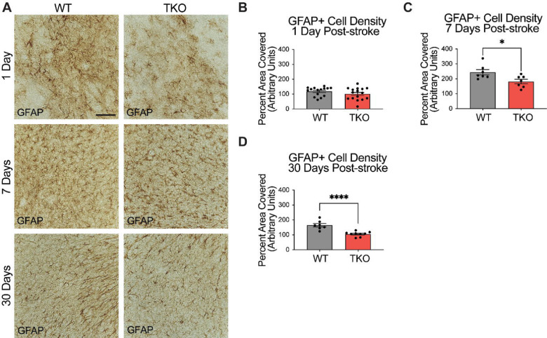
Figure 2. TNF, IL1α, and C1q knockout reduces GFAP+ cell number in the peri-infarct area 7- and 30-days post ischemic stroke.
(A) Representative images of GFAP+ cells in the peri-infarct area for wildtype (WT) and Tnf−/−Il1a−/−C1qa−/− triple knockout (TKO) mice 1-, 7-, and 30-days post ischemia. (B) No difference was found in GFAP+ cells in the peri-infarct area 1 day after ischemia between the WT and TKO mice, t (29) = 1.29, p = .104. (C) TKO mice had significantly reduced GFAP+ cells in the peri-infarct area than WT mice 7 days post-stroke, t (12) = 2.64, p = .011. (D) TKO mice had significantly reduced GFAP+ cells in the peri-infarct area than WT mice 30 days post-stroke, t (14) = 5.03, p < .0001. * p < .05, ** p < .001, *** p < .0001. Abbreviations: GFAP; glial fibrillary acidic protein, TKO, Tnf−/−Il1a−/−C1qa−/− triple knockout mouse; WT, wild type.

