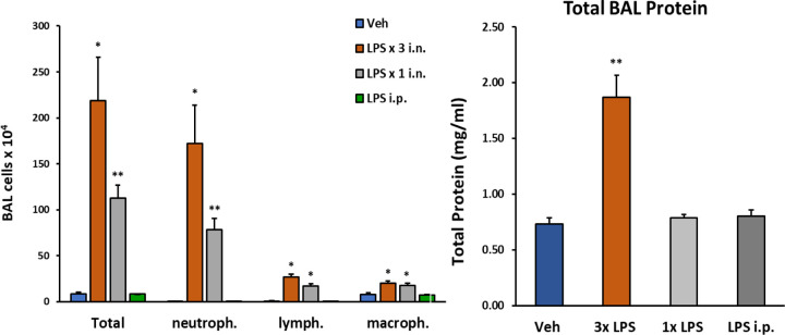
Figure 1: Changes in lung BAL following LPS exposures.
C57/Bl6 male mice (12 weeks old) received either 1 i.n. (10μg/40μl), 3 i.n (30μg/40μl). or 1 i.p. (2mg/kg) administrations of LPS from Escherichia coli O111:B4 purified by phenol extraction (Sigma, L2630) in sterile saline (Veh). Mice were sacrificed 24h after last LPS dose (or after single LPS i.n. or i.p). Number of cells detected in BAL fluid was significantly increased following 3 i.n. LPS exposures/daily; total protein content in BAL was also robustly increased following this protocol. Data analyzed with student t-test: p<0.05(*), p<0.01(**), p<0.001(***), p<0.0001(****), data represented as mean (SEM).

