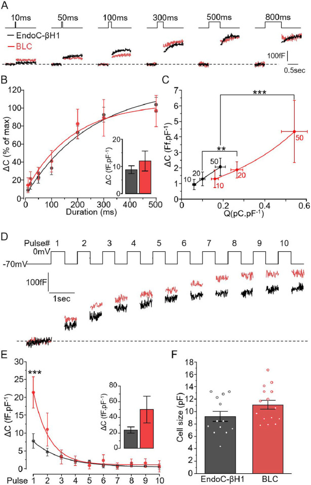
Figure 3: Comparison of the exocytotic properties of BLCs and EndoC-βH1 cells.
A-B. EndoC-βH1 (black) and BLCs dispersed from cluster preparations (red) were subjected to increased durations of depolarisation (from 10 ms to 800 ms) and the resulting exocytosis events were measured as an increase in cell surface area. B. The stimulations triggered similar kinetics of exocytosis in both models with a plateau reached from 300ms depolarisation. Inset: maximum exocytosis elicited by 800ms stimulation (n=13 and n=15 cells for EndoC-βh1, black, and BLC, red, respectively). C. Charge-exocytosis relationship in EndoC-βH1 and in BLC. The right-shifted correlation in BLCs is driven significantly by the amount charges. D. Representative traces of the maximum exocytosis elicited by ten depolarisations of 500 ms. F. Quantification of exocytosis increment elicited at each pulse (n=13 and 11 cells for EndoC-βH1 and BLC, respectively). Insert: total cumulative exocytosis measured at pulse 10. G. The initial size of the cells (pF) were similar between the two β-cell models (n=13 and n=15 cells for EndoC-βh1, black, and BLC, red, respectively). Statistics, **p<0.01, *** p<0.001, Paired comparison and Bonferroni posthoc test.

