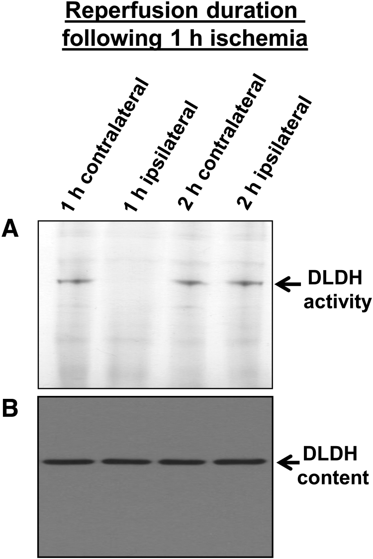
FIG. 6.
BN-PAGE measurement of DLDH diaphorase activities (A) and Western blot assay of DLDH protein content (B) after ischemic stroke. Rat brain underwent 1 h ischemia followed by 1 or 2 h reperfusion. As can be seen in this figure, upon a 1-h reperfusion, no DLDH activity could be detected (A, lane 2 from the left), but protein content did not change (B). However, DLDH activity was fully recovered after a 2-h reperfusion. This figure was reproduced with copyright permission from reference (Wu et al., 2017).

