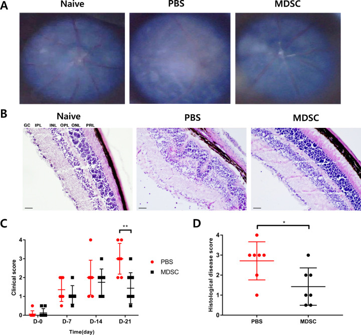
Figure 2.
Clinical scoring and histopathological grading of the retina following the administration of MDSCs in EAU. (A) Fundus images of mice showing representative photos in each group. (B) Histologic representative images indicating retinal tissues stained with hematoxylin and eosin in each group. Scale bars, 50 µm. GCL, ganglion cell layer; IPL, inner plexiform layer; INL, inner nuclear layer; OPL, outer plexiform layer; ONL, outer nuclear layer; PRL, photoreceptor layer. (C) Locally injected MDSC group (n = 8) displayed significantly less intraocular inflammatory scores than the PBS group (n = 7) for 21 days (**P = 0.0027). (D) The histopathological grading of EAU showed a significant decrease in the MDSC group (n = 7) compared with that in the PBS group (n = 8) (*P = 0.0253). Data are represented as mean ± SD of three independent experiments.

