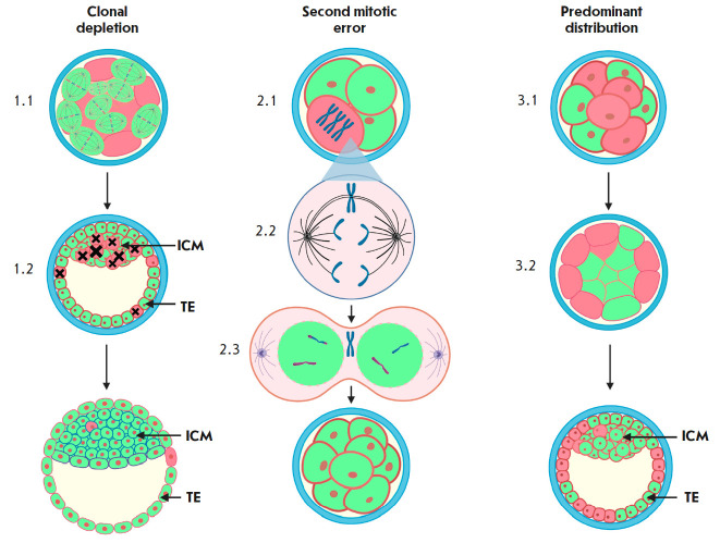
Fig. 2.
Models of self-correction of the chromosomal status in mosaic embryos. Euploid cells are indicated in green, aneuploid cells are indicated in pink. Spindles (1.1) reflect an increase in the proliferative activity of euploid cell lines in mosaic embryos. Black crosses indicate apoptotic processes in aneuploid cells (1.2). A trisomal aneuploid embryonic cell (2.1) can undergo corrective mitotic division. One of the chromosomes remains at the mitotic spindle equator due to merotelic attachment of microtubules to kinetochores (2.2) and is not further included in the nuclei of daughter cells (2.3). (3.1, 3.2) Displacement of aneuploid cells to the embryo periphery, to the area of the nascent trophectoderm

