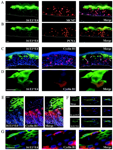
FIG. 7.
Expression of 16E1∧E4 in cyclin B1-expressing cells during epithelial cell differentiation. HPV16-induced raft cultures were double stained for the presence of 16E1∧E4 (green) and (A) MCM7, (B) PCNA, or (C) cyclin B1 (red) to show that 16E1∧E4 levels begin to rise in cells that are expressing cell cycle markers. A subset of the cells that express moderate levels of 16E1∧E4 also express cyclin B1 and are arrowed in C. (D) An enlarged image of the cyclin B1/16 E1∧E4 double-positive cell that is marked with an asterisk in C. (E) HPV16-induced low-grade lesion double stained for the presence of 16E1∧E4 (green) and cyclin B1 (red). Coexpression of cyclin B1 and 16 E1∧E4 is limited to the region wherethe levels of 16E1∧E4 protein are beginning to rise during epithelial differentiation. Panel G shows a high-power image of cells from this region (indicated by an asterisk in E) that contain both cyclin B1 and 16 E1∧E4. Panel F shows that 16E1∧E4-expressing cells (green) in the upper layers of low-grade cervical lesions contain the keratin phosphoserine 33 epitope (red) that is also present following the expression of 16E1∧E4 in vitro (see Fig. 5). Merged images are shown in the uppermost panels within F. Where appropriate, the position of the basal layer is indicated by dotted white lines. A DAPI nuclear stain (blue) was included. Bars equal 20 μm except in panels D and G, where the bars equal 10 μm.

