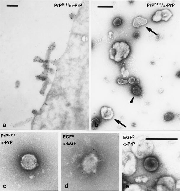
FIG. 4.
Immunoelectron microscopic analysis of the particles. Sections through HEK-293FT producer cells transfected with pHIT60/pD-PrP111 (a) and PrPD111 retroparticles harvested from the cell culture supernatant and concentrated by low-speed centrifugation (b and c) were stained with the PrP-specific 6H4 antibody and a 10-nm-diameter gold particle-labeled anti-mouse IgG. EGF retroparticles (d and e) were harvested from pHIT60/pD-EGF-transfected 293FT cells and stained with the anti-EGF (d) or the 6H4 (e) antibody. Bars, 250 nm. Arrowheads indicate particles with the typical morphology of C-type retroviruses, and arrows indicate pleomorphic vesicles.

