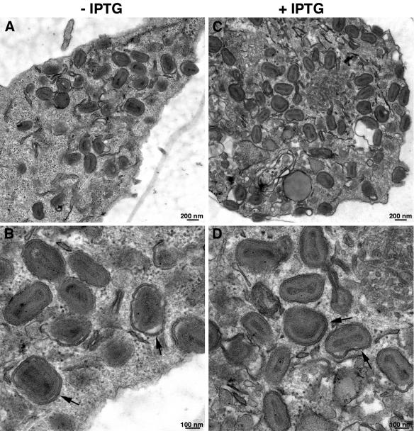
FIG. 6.
Effect of GFP-EH21 on formation of IEV and CEV. BS-C-1 cells were infected with vEH21i for 8 h in the absence (A and B) or presence (C and D) of IPTG. Thin sections were viewed by transmission electron microscopy, and images were obtained at low (A and C) and high (B and D) magnifications. The arrows point to the representative IEV membranes. The magnifications are indicated by the scale bars.

