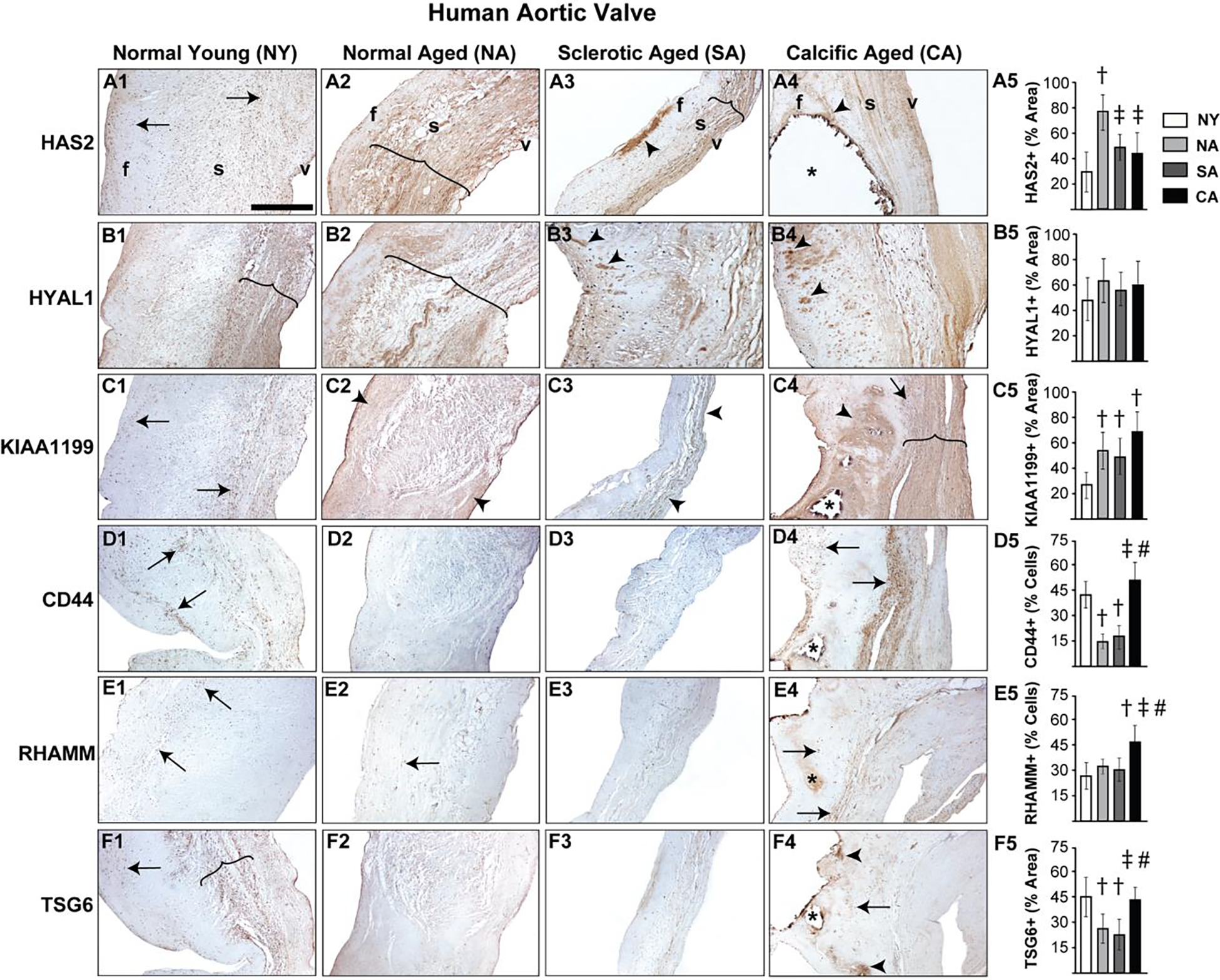
Figure 1.

Diseased human aortic valves demonstrate abnormalities in regional expression of HA homeostatic markers. Expression patterns of HAS2 (A1-A5), HYAL1 (B1-B5), KIAA1199 (C1-C5), CD44 (D1-D5), RHAMM (E1-E5) and TSG6 (F1-F5) in valves classified as young (15–20 year old) or aged (>50 year old) and either normal or abnormal (sclerotic/calcific) are shown. Arrows and arrowheads denote cell and ECM expression respectively. The region of expression is marked by parenthesis. Calcific nodules are indicated by asterisk. †: vs normal young p<0.05; ‡: vs normal aged p<0.05; #: vs sclerotic aged p<0.05. f, fibrosa; s, spongiosa; v, ventricularis. The orientation provided by the use of f, s, v in panels A1-A4 apply to all the microscopic image panels in this figure. Scale bar: 200 μm.
