Figure 7.

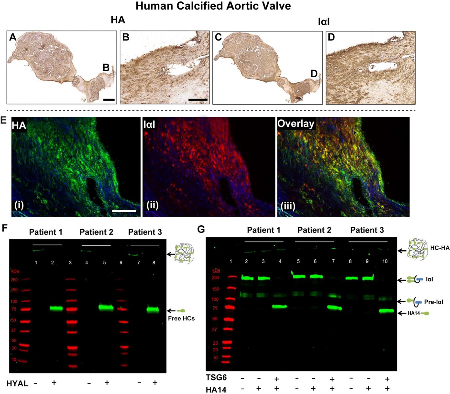
Human calcified aortic valves demonstrate extensive HA and IαI expression, and their co-localization (A-E). Their secondary-only controls are included in Supplemental Fig 5 for comparison. F) Western blot demonstrating presence of the pathological form of HA (i.e., heavy chain (HC)-HA complex) in these valves. When hyaluronidase (HYAL) is added to the minced tissue, HC-HA, which is too large to enter the gel, is digested, releasing free HCs that migrate and appear as a single band at ~85 kDa (green bands; lanes 2, 5, 8). G) The TSG6 activity assay works by supplying IαI (endogenous, from the tissue) and a HA oligo (exogenous) 14 monosaccharides in length (HA14) as an artificial heavy chain acceptor. If endogenous TSG6 activity was present, it would have transferred the HCs from IαI to HA14, and there would have been a gel shift from the 250 kDa IαI band to the ~85 kDa HC band in lanes 3, 6 and 9. This was demonstrated in the positive controls where recombinant TSG6 was supplied (green bands; lanes 4, 7, 10). TSG6 activity was largely absent in the calcified human aortic valves. Molecular weight standards are shown in red in both F and G. Scale bar: A, C: 0.5 mm; B, D, E: 200 μm.
