
Figure 8.
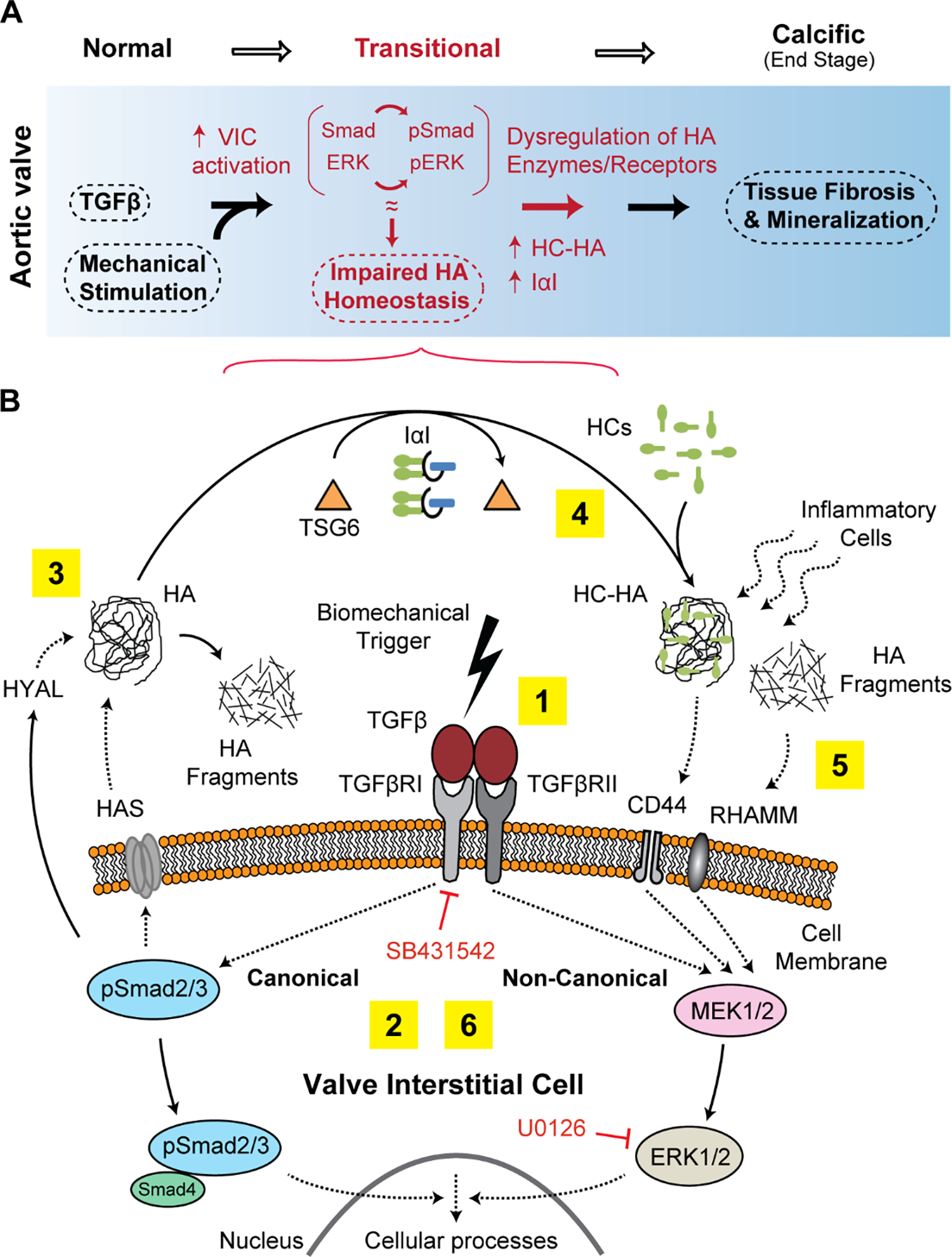
Schematic mechanism of the association between TGFβ signaling, biomechanics and HA homeostasis in calcific AVD progression. A) We propose that activation of intracellular TGFβ pathways together with biomechanical trigger cause a transitional, pre-calcific phase (red markings) with increased cell activation, ECM remodeling and dysregulation of enzymes that synthesize and breakdown HA. B) HA abnormalities manifesting as either accumulation of HA fragments (due to increased HYAL, denoted by 3) or HC-HA (due to increased TSG6 activity, denoted by 4) may impair normal HA receptor function and may attract inflammatory cells to the site of calcific lesions (denoted by 5). The numbers 1 through 6 denote the proposed sequence of events; dotted arrow between the moieties means “acts on”, and solid arrow means “converts to”.