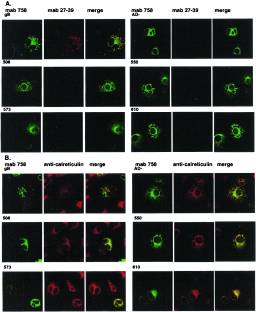
FIG. 7.
gB cysteine mutants fail to exit the ER and are not recognized by oligomer-specific MAb 27-39. Wild-type gB and gB mutants ΔgBAD-1 (designated AD− in this figure), ΔgBc508s, ΔgBc550s, ΔgBc573s, and ΔgBc610s were transiently expressed in COS-7 cells grown on glass coverslips and fixed at 48 h in 3% paraformaldehyde. (A) The cells were stained with MAb 758, the oligomer-specific MAb 27-39, and developed with an FITC-labeled goat anti-human IgG or Texas red-conjugated goat anti-mouse IgG second antibody. Images were merged to determine colocalization. (B) The cells were stained with MAb 58-15 or with rabbit anti-calreticulin and developed with FITC-conjugated goat anti-mouse IgG or Texas red-labeled anti-rabbit IgG. Images were merged to determine colocalization.

