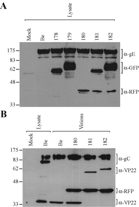
FIG. 1.
Expression and virion incorporation of fluorescent fusion proteins. PK15 cells were either mock infected or infected with the indicated PRV strains at an MOI of 10 for 16 h prior to preparation of whole-cell lysates or purified extracellular virions (as indicated). Western blot analysis was performed using monoclonal antibodies to gE, GFP, and RFP (A) or polyclonal antibodies to gC, VP22, and RFP (B). The migration positions of molecular mass markers are shown on the left (in kilodaltons). Be, Becker strain.

