FIG. 3.
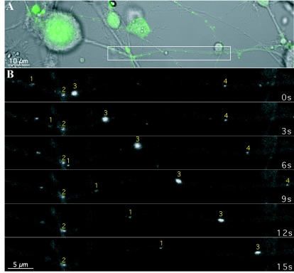
Transport of GFP-VP22 structures in the axonlike projections of differentiated rat PC12 cells. PC12 cells differentiated with NGF were infected with PRV 178 at an MOI of 10 for approximately 12 h prior to live-cell confocal microscopy in a heat-controlled environment. (A) The cell bodies and projections were visualized by differential interference contrast microscopy while the GFP autofluorescence, indicative of infection, was readily detectable by confocal microscopy. (B) A section of the projection (inset in panel A) was imaged by time-lapse microscopy. Several GFP puncta of varying emission intensity are visible. The transport properties of these structures during the imaging sequence also varied; two were moving (puncta 1 and 3), one was briefly stationary and then moved (punctum 4), and another remained stationary (punctum 2).

