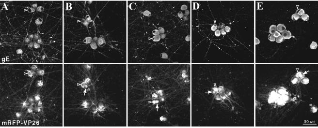
FIG. 5.
Time course of BFA treatment during infection. Dissociated rat SCG neurons were infected with PRV 180 at a high MOI for 15 h prior to fixation and detection of gE localization by indirect immunofluorescence and mRFP-VP26 autofluorescence. Infected neurons were untreated (A) or subjected to a concentration of 1 μg of BFA/ml from 12 (B), 9 (C), 6 (D), or 3 (E) h postinfection until the time of fixation. Examples of nuclei of BFA-treated cells are indicated with hollow arrowheads (A to E). Cytoplasmic accumulations of fluorescent capsid are indicated with arrows (B to E).

