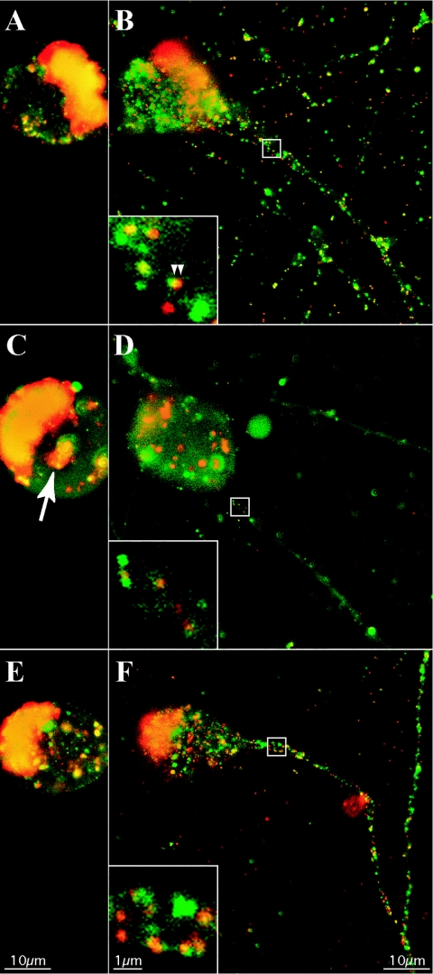
FIG. 7.
Prolonged BFA treatment dramatically reduces the axonal entry of fluorescent capsid and tegument puncta. Dissociated rat SCG neurons were infected with PRV 181 at a high MOI and then subjected to incubation for 12 h (A and B), treatment with 2 μg of BFA/ml from 2 to 12 h postinfection (C and D), or treatment with BFA as before with recovery from 12 to 18 h postinfection (E and F) prior to detection of autofluorescence by confocal microscopy. Two planes of focus are shown: through the center of the nucleus, above axons (A, C, and E), or below the center of the cell body, through axons (B, D, and F). A region of axon containing fluorescent puncta (white box) is shown at a higher magnification (inset, lower left) (B, D, and F), while a cytoplasmic accumulation of fluorescent capsid and tegument is indicated with an arrow (C).

