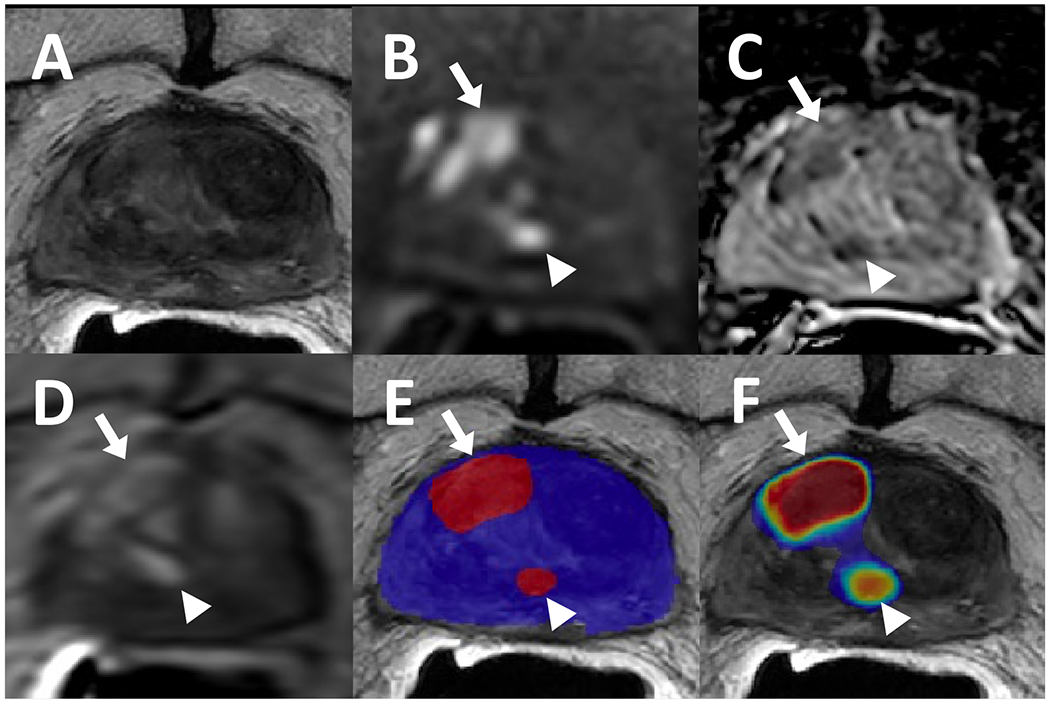
Figure 3.

82-year-old male with a history of external beam radiation therapy for prostate cancer and serum PSA level of 9.33 ng/ml. The patient had lesions in right mid-base anterior transition zone (arrows) and right mid periurethral transition zone (arrowhead) which were visible on T2-weighted imaging (A), ADC map (B), high b-value diffusion weighted imaging (DWI) (C), and dynamic contrast enhancement (DCE) (D). Targeted biopsies obtained from these sites were positive for recurrent cancer, and both were detected by the AI model as displayed in a binary AI prediction map (E) and a probability map (F).
