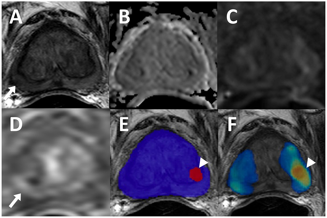
Figure 6.

A 72-year-old male with a history of external beam radiation therapy for prostate cancer and serum PSA level of <0.01 ng/ml. Patient had a lesion in right mid peripheral zone which appears slightly hypointense on T2-weighted imaging (A) (arrow), negative on ADC map (B) and high b-value diffusion weighted imaging (DWI) (C) with a mild focal contrast uptake on dynamic contrast enhancement (DCE) (D) (arrow). Targeted biopsy revealed residual cancer, however, AI model did not detect this lesion. In left mid transition zone, the AI model had a false positive prediction, visible on binary AI prediction map (E) and probability map (F) (arrowhead) which may be attributed to the signal abnormalities caused by an adjacent fiducial marker.
