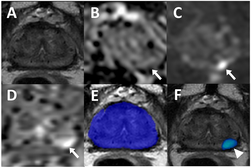
Figure 8.

A 70-year-old male who received brachytherapy for prostate cancer treatment with a serum PSA level of 1.5 ng/ml. Patient has a lesion in left apical-mid peripheral zone which is not visible on T2-weighted imaging (A) but appears hypointense on ADC map (B) and hyperintense on high b-value diffusion weighted imaging (DWI) (C) (arrows). The lesion also demonstrates early enhancement on dynamic contrast enhancement (DCE) (D) (arrow). Recurrent prostate adenocarcinoma was confirmed on targeted biopsy. Biparametric MRI-based AI did not have any prediction on the binary AI prediction map (E) and therefore, this was considered a false negative, however, the probability map (F) localized the cancerous focus (arrowhead).
