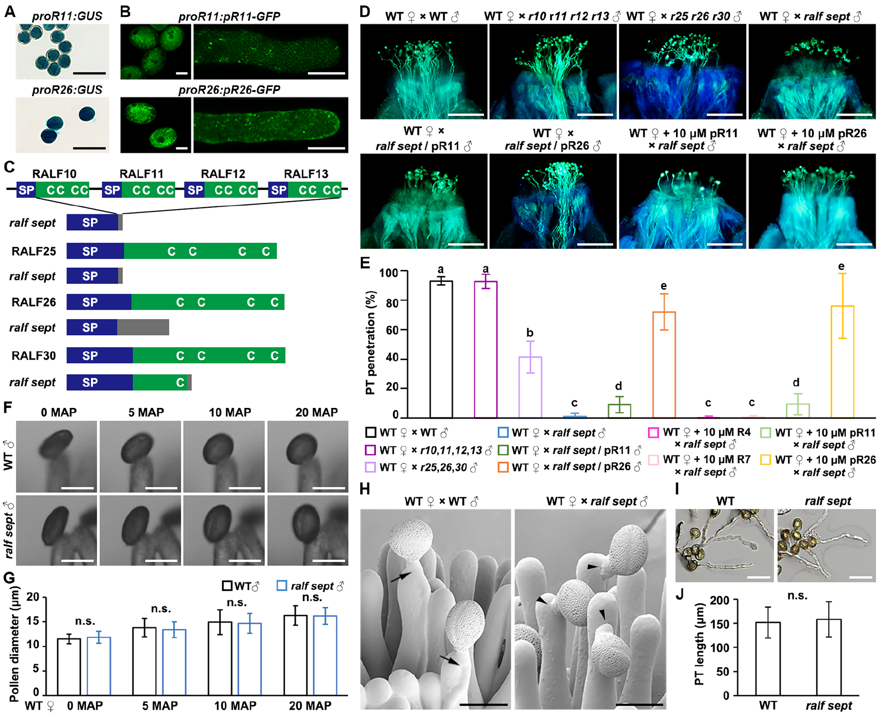
Figure 1. Seven pollen-specific RALF peptides (pRALFs) promote pollen-tube penetration in Arabidopsis.

(A) GUS staining images show promoter activities of pRALF11 and pRALF26 (abbreviated as pR11 and pR26, respectively) in pollen. Scale bars, 50 μm.
(B) Mature pollen grains and pollen tubes expressing protein fusions of pR11/pR26-GFP driven by their respective native promoters. Scale bars, 10 μm.
(C) Schematic diagrams describe specific mutations created by CRISPR/Cas9-edited mutagenesis in the ralf10 ralf11 ralf12 ralf13 ralf25 ralf26 ralf30 septuple mutant (abbreviated as ralf sept). Native peptide structures were aligned above respective mutations. C, conserved cysteine residues; SP, signal peptide. Gray boxes indicate missense sequences due to frame shift or fragment deletion.
(D) Aniline blue staining shows status of pollen tube penetration in the wild-type (WT) stigmas at 3 hours after pollination (HAP). Pollen applied include WT, r10 r11 r12 r13, r25 r26 r30, ralf sept, pR11-complemented ralf sept, or pR26-complemented ralf sept. +10 μM pR11/26 indicate WT stigmas that were pre-treated with synthetic pR11 or pR26 peptides, respectively. Scale bars, 200 μm.
(E) Statistical analysis of pollen tube penetration assayed in (D) with negative controls shown in Figure S2J. Experiments were repeated at least three times. Different letters represent significant differences between groups (P<0.001).
(F) Representative bright field images show pollination-induced pollen hydration of WT or ralf sept pollen on WT stigmas. MAP, minutes after pollination. Scale bars, 20 μm.
(G) Measurement and statistical analysis of pollen diameter during pollen hydration in (F). Equatorial diameters of pollen grains were measured.
(H) Cryo-Scanning Electron Microscopic (CSEM) images of WT stigmas pollinated with WT or ralf sept pollen at 3 HAP. Black arrows point to WT pollen tubes penetrating the papilla cells and black arrow heads point to ralf sept pollen tubes failing to penetrate the papilla cells. Scale bars, 25 μm.
(I) In vitro growth of WT and ralf sept pollen tubes. Images were collected after incubation for 6 hours. Scale bars, 50 μm.
(J) Statistical analysis of pollen tube length in (I). Experiments were repeated at least three times. N>100 for each group. Data in (E, G and J) are mean values ± SD. n.s., not significant, P>0.1 (Student’s t test).
