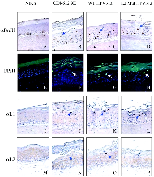
FIG. 2.
Immunohistochemical analyses of rafts. (A to D) Immunohistochemistry for BrdU incorporation. (E to H) FISH with fluorescein isothiocyanate-conjugated antibody specific for DIG-labeled HPV31 DNA probe. (I to L) Immunohistochemistry for L1 expression with H16.D9 antibody. (M to P) Immunohistochemistry for L2 expression with αHPV31-L2 antibody. For all panels displaying immunohistochemical staining (A to D and I to P), brown cells are positive and are indicated with blue arrows. Counterstain was done with hematoxylin. For all panels displaying immunofluorescent staining (E to H), cells positive for FISH signal have green nuclei (white arrows), whereas FISH-negative cells have blue nuclei (a consequence of the DAPI counterstain). Sections present in the first column (A, E, I, and M) are taken from the negative control NIKS rafts, those in the second column (B, F, J, and N) are from the positive control CIN-612 9E rafts, those in the third column (C, G, K, and O) are from wild-type (WT) HPV31a-harboring rafts, and those in the fourth column (D, H, L, and P) are from L2 mutant HPV31a-harboring rafts.

