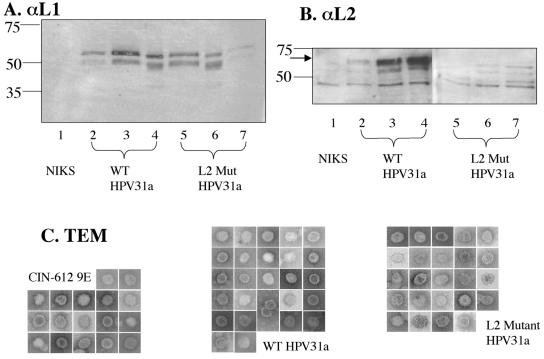
FIG. 4.
Analysis of crude preparations of virus. (A and B) Immunoblot (Western) analyses of equal volumes of crude virus preparations from rafts of three different clonal NIKS populations each harboring wild-type (WT; lanes 2 to 4) or L2 mutant (L2 Mut; lanes 5 to 7) HPV31a genomes, along with a sample prepared in parallel from rafts of the negative control (untransfected) NIKS cell line (lane 1). Numbers at left indicate molecular mass in kilodaltons. In panel A, L1 was detected using the monoclonal antibody H16.D9. Note the lower levels of L1 detected in lanes 2 and 7, which reflect these particular samples yielding less virus. In panel B, L2 was detected using a polyclonal antibody to HPV31 L2. The arrow indicates the L2-specific band. Note the presence of multiple nonspecific bands that migrated faster than the L2-specific band and are present in the negative control sample isolated from rafts of untransfected NIKS (lane 1). (C) TEMs of particles of about 55 nm in diameter. The side of each square is 100 nm long. Images are shown at 110,00× magnification. No particles of 55 nm in diameter were seen in the crude preps of NIKS cells.

