Figure 4.

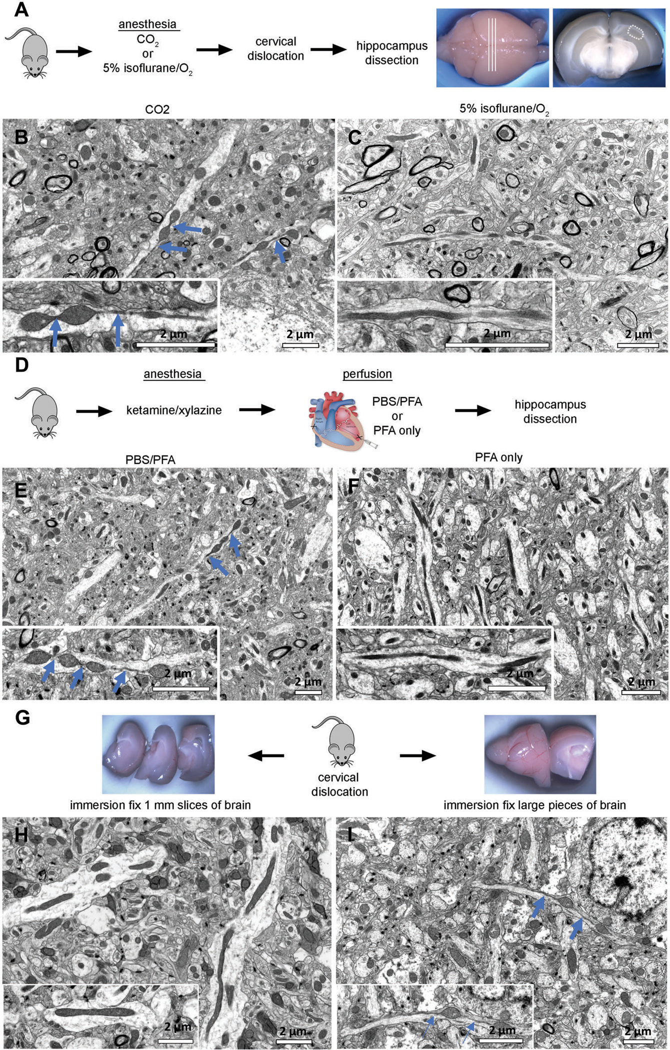
Evaluation of conditions to reduce artifacts in tissue preparation to assay mitochondrial morphology using EM. A) Wild-type (WT) mice were sacrificed by cervical dislocation after anesthesia with CO2 or 5% isoflurane/oxygen inhalation. Fresh brains were removed and cut into coronal sections. The CA1 hippocampal region (hipp) from each section was dissected and further processed for TEM or scanning electron microscopy (SEM). B) CO2 exposure for 3 min was sufficient to induce MOAS formation in hippocampal tissue of WT mice. Scale bar: 2 μm. C) Mitochondria in hippocampus from mice administered 5% isoflurane prior to cervical dislocation and subsequent dissection maintained a normal shape and size typical for WT mice. Scale bar: 2 μm. D) WT mice were anesthetized using ketamine/xylazine, followed by cardiac perfusion with either phosphate-buffered saline (PBS) an 4% paraformaldehyde (PFA) or 4% PFA only. Mice were euthanized by cervical dislocation, and brain removed and fixed in Trump’s solution overnight. The next day, brains were cut into coronal sections. The CA1 hippocampal region (hipp) from each section was dissected as shown in (A) and subjected to further processing for TEM or SEM. E,F) Cardiac perfusion preceded by PBS flush induced MOAS in hippocampal tissue of WT mice. Perfusion without PBS flush did not cause MOAS. Scale bar: 2 μm. G) WT mice were euthanized by cervical dislocation without prior anesthesia. Brains were removed, and one hemisphere was cut into 1-mm-thick slices (left), while the other hemisphere was cut into two halves (right). Tissues were subjected to immersion fixation in Trump’s solution overnight. H) Tissues cut in thin slices were free of MOAS, I) while larger pieces of tissue displayed pronounced MOAS. Scale bars: 2 μm. Blue arrows indicate MOAS.
