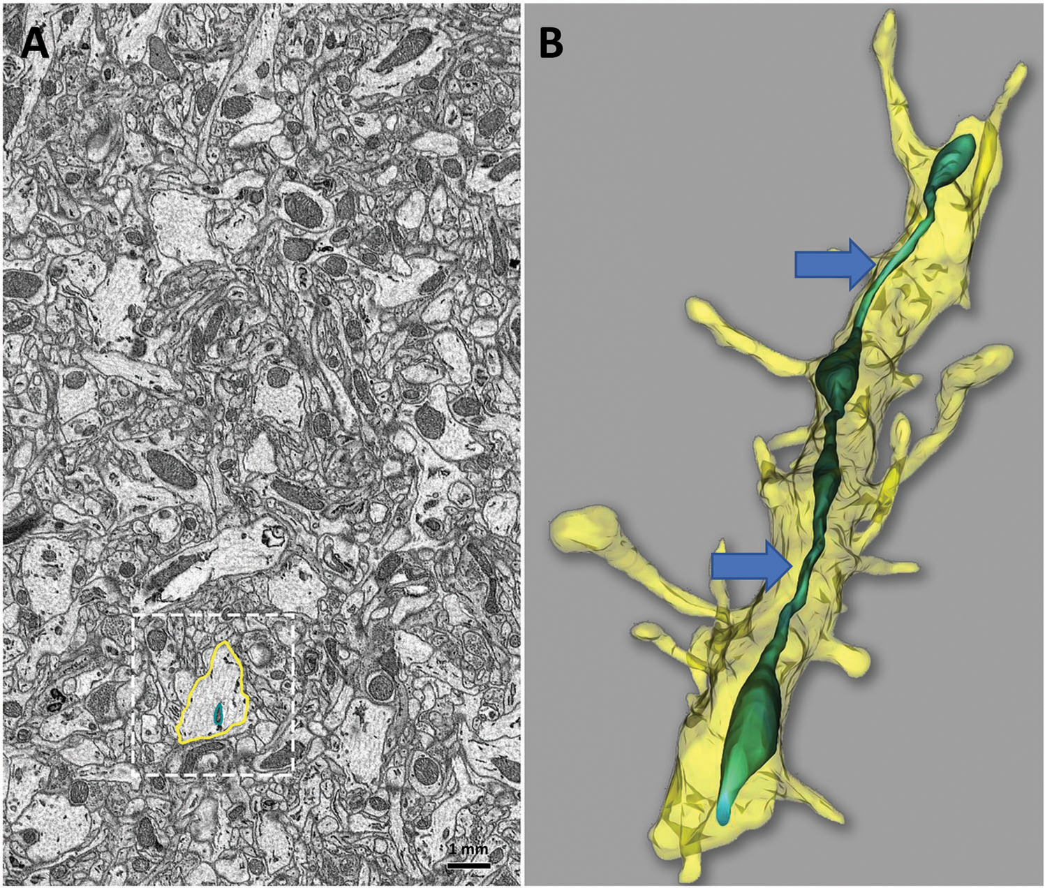
Figure 5.

Serial sectioning and 3D EM reconstruction provide additional insight into ultrastructural details of mouse brain tissue that cannot be fully observed in 2D. A) Minimal details of a single neuropil and mitochondria are observed in a 2D micrograph of mouse hippocampal tissue (A, boxed area). B) 3D EM reconstruction of the same brain area using serial block-face imaging allows identifica neuropil as a dendrite, based on the presence of dendritic spines and reveals mitochondrial ultrastructure as MOAS (blue arrows). Reconstruct software was used for 3D reconstruction. Scale bar: 1 mm.
