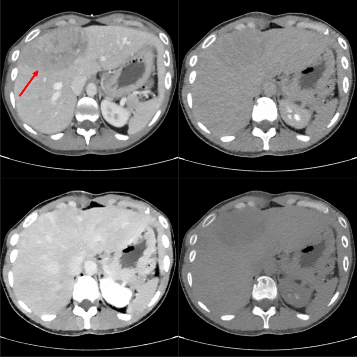
Fig. 3.
Patient #7, the shown lesion was histopathologically identified as a breast cancer metastasis. Shown are the diagnostic CT in the venous phase (top left), as well as the corresponding biopsy images. The lesion is best visible in the VNC image (bottom right) due to hypodensity compared to the remaining liver parenchyma. It is also visibility in the conventional reconstruction (top right) and worst visible in the VMI-40 image (bottom left)

