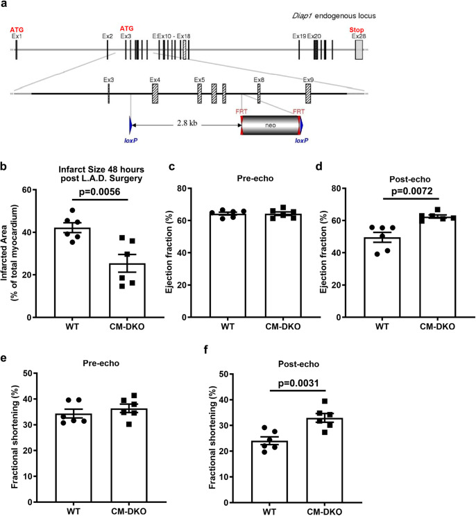
Fig. 5. Deletion of CM-specific Diaph1 is protective in ischemia/reperfusion injury.
a Generation of Diaph1 floxed mice. Schematic representation of Diaph1 targeting strategy, resulting in deletion of exons 4–7. The diagram is not depicted to scale. Hatched rectangles represent Diaph1 coding sequences, gray rectangles indicate non-coding exon portions, and solid line represents chromosome sequence. The initiation codon (ATG) and stop codon (Stop) are indicated. FRT sites are represented by double red triangles, and loxP sites by blue triangles. Diaph1flox/flox Cre (+) (“CM-DKO”) and Diaph1flox/flox Cre (−) (“WT”) mice hearts which underwent LAD ligation for 30 min creating ischemic/no flow condition followed by reperfusion to mimic I/R injury. b Quantification of infarct size in CM-DKO and WT hearts (n = 6 biologically independent samples, unpaired t-test was performed for p value). 2D-echocardiography measurements of (c, d), ejection fraction (n = 6 biologically independent samples, unpaired t-test was performed for p value) and e, f fractional shortening 48 h post LAD surgery in CM-DKO and WT mice (n = 6 biologically independent samples, Wilcoxon rank-sum test was performed for pre-echo and unpaired t-test was performed for post-echo p value). Data are presented as the mean ± SEM. All statistics and source data are provided as a Source Data file.

