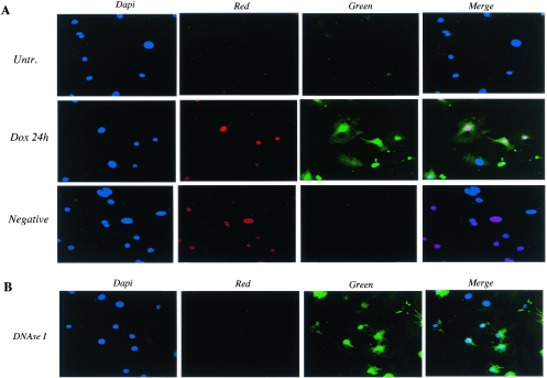
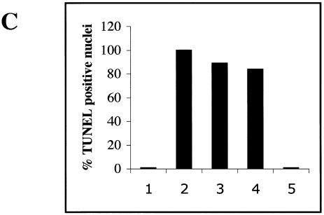
FIG. 9.
p300 degradation in cardiomyocytes treated with doxorubicin parallels apoptosis. (A) Primary neonatal cardiomyocytes plated on coverslides were maintained in normal medium (Untr.) or in medium supplemented with 1 μM doxorubicin for 24 h (Dox 24 h) and were subjected to the TUNEL assay as described in Materials and Methods. The cells were then mounted with DAPI-containing medium to visualize the nuclei. Apoptotic cells were analyzed by confocal microscopy and were detected by fluorescein (Green). Nuclei stained in red represent doxorubicin intercalation in the nuclei of cardiomyocytes (Red). (B) For a positive control, we used cardiomyocytes permeabilized and then incubated with DNase 1 to induce DNA strand breaks. (C) Percentage of apoptotic nuclei in untreated cells (bar 1), in DNase I-treated cells (bar 2), in cells treated for 15 h (bar 3) and 24 h (bar 4), with doxorubicin and in the negative control (bar 5).


