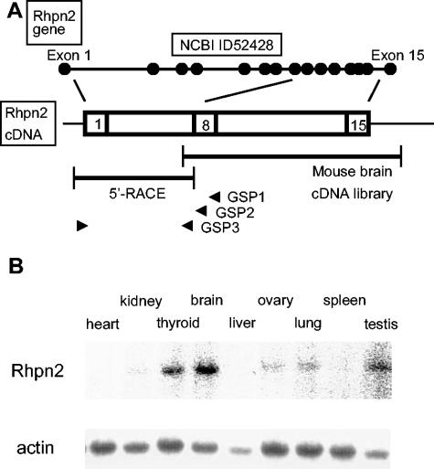
FIG. 1.
Mouse rhophilin 2 gene structure, cDNA, and expression in tissues. (A) Schematic representation of the exon-intron structure of the mouse rhophilin 2 gene on chromosome 7, as reported in NCBI databases (see above). The two DNA fragments isolated by screening of a mouse brain cDNA library and by 5′ RACE are depicted as well as the three specific primers (GSP1, GSP2, and GSP3, all located in exon 8 [arrowheads]) used in 5′ RACE (see below). (B) Hybridization analysis of RNA isolated from mouse tissues with a rhophilin 2 probe or an actin probe.

