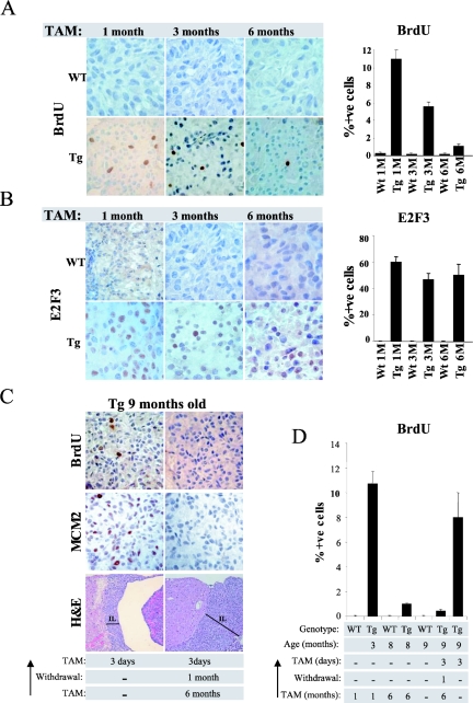
FIG. 3.
Melanotrophs become resistant to sustained increased E2F activity. (A) Decreased proliferation following sustained E2F activation. Bromodeoxyuridine incorporation measured by immunohistochemistry in the intermediate lobe of wild-type (WT) and transgenic (Tg) animals treated for the indicated time with daily tamoxifen (TAM) injections. The bar graph indicates the percentage of bromodeoxyuridine-positive melanotrophs in the intermediate lobe and the standard deviation of the mean. (B) Expression of ER-E2F3 protein is retained in tamoxifen-treated mice. Immunohistochemistry analysis of ER-E2F3 expression in the intermediate lobe of wild-type (WT) and transgenic (Tg) animals treated for the indicated times with daily tamoxifen injections. The bar graph indicates the percentage of E2F3-positive melanotrophs in the intermediate lobe and the standard deviation of the mean. (C) Sustained E2F activity triggers melanotrophs to become refractory to E2F stimulation. From the top: bromodeoxyuridine incorporation, MCM2 expression, and hematoxylin and eosin staining of melanotrophs after 3 days of tamoxifen treatment of 9-month-old transgenic mice either not previously exposed to tamoxifen (left panel) or treated for 6 months followed by 1 month of withdrawal (right panel). Note that the hyperplastic morphology of the intermediate lobe (bar) of the long-term-induced transgenic animals does not regress upon tamoxifen withdrawal (right panel). (D) Percentage of bromodeoxyuridine-positive melanotrophs. Treatment of 9-month-old transgenic animals leads to the same extent of S-phase induction achieved in 3-month-old transgenic animals (compare second lane from left with last on the right). Sustained E2F activity prevents S-phase induction in melanotrophs (fourth and sixth lanes from the left).

