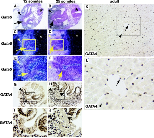
FIG. 5.
Expression of GATA4 but not GATA6 is extinguished in migrating hepatoblasts. Embryos containing 12 (A, C, E, G, and I) or 25 (B, D, F, H, and J) somite pairs and adult livers (K and L) were isolated and processed for in situ hybridization to detect Gata6 mRNA (A to F) or for immunohistochemistry to detect GATA4 protein (G to L). Both GATA4 protein (brown nuclear staining; G and I) and Gata6 mRNA (A, C, and E) can be detected in the septum transversum mesenchyme (arrowheads) and liver bud (arrows) of 12-somite-pair embryos. At 25 somite pairs, Gata6 mRNA and GATA4 protein were detected in septum transversum mesenchyme (arrowheads; B, D, F, H, and J). In the liver bud (arrows; B, D, F, H, and J), however, the presence of Gata6 mRNA could be identified but GATA4 protein was undetectable. GATA4 protein was also detected predominantly in the endothelial cells (arrows) but not in hepatocytes (arrowheads) of adult livers (K and L). Panels E, F, I, J, and L show high-resolution images of boxed areas in panels C, D, G, H, and K, respectively. Both bright-field (A and B) and corresponding dark-field (C to F) images of Gata6 in situ hybridization analyses are presented. The presence of mRNA can be detected as bright silver grains in dark-field images; note the absence of staining in neural tubes (asterisks).

