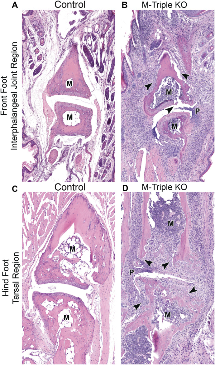
Figure 3. Paw histology.
(A, B) H&E-stained sections of the interphalangeal joint region of front paws from (A) male control and (B) male M-triple KO mice at 7–9 wk of age (20X magnification). (C, D) H&E-stained sections of the metatarsal–tarsal region of the hind foot from (C) male control and (D) male M-triple KO mice at 7–9 wk of age (20X magnification). Arrowheads in the M-triple KO sections indicate neutrophilic infiltration into the bone, cartilage, and soft tissue. Note: marrow cavity (M) is full of myeloid cells in the M-triple KO sections, compared with an essentially empty marrow cavity (M) in the control sections. In the M-triple KO paw sections, the synovium or pannus (P) is protruding into the joint space.

