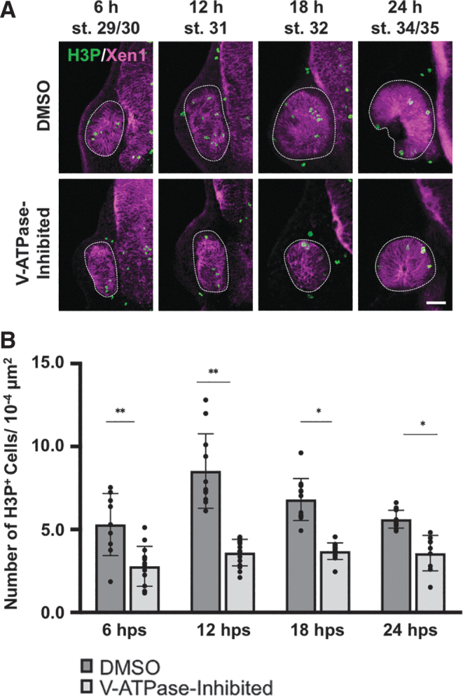
FIG. 3.

V-ATPase regulates cell proliferation in regrowing eyes. (A) Representative fluorescence images of eye sections stained with anti-H3P antibody to identify mitotic cells (green). Magenta color indicates neural tissues (Xen1). White dashed lines outline each eye. Sample size ranges from n = 9 to 12 per condition and timepoint. Up = dorsal. Scale bar = 50 μm. (B) Quantification of mitoses in control DMSO regrowing and V-ATPase−inhibited eyes. Graph shows the number of H3P-positive cells. *P < 0.05, **P < 0.01 (n = ≥9 per timepoint and condition). Data are means ± SD.
