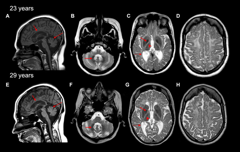
Figure 1.
Brain MRI characteristics of the affected individual at 23 and 29 years old. A thin corpus callosum (red arrow) and cerebellar vermis atrophy (red dashed arrow) are seen on sagittal T1-weighted MRI images (A,E). Cerebellar hemisphere atrophy was also seen at 23 and 29 years (not shown). Diffuse hypomyelination is evident (C,D,G,H), with relative preservation of myelin seen in the dentate nucleus (red arrow in B,F), globus pallidus (red arrow in C,G), the anterolateral nucleus of the thalamus (red arrowhead in C,G), and optic radiations (dashed red arrow in C,G) on axial T2-weighted MRI images.

