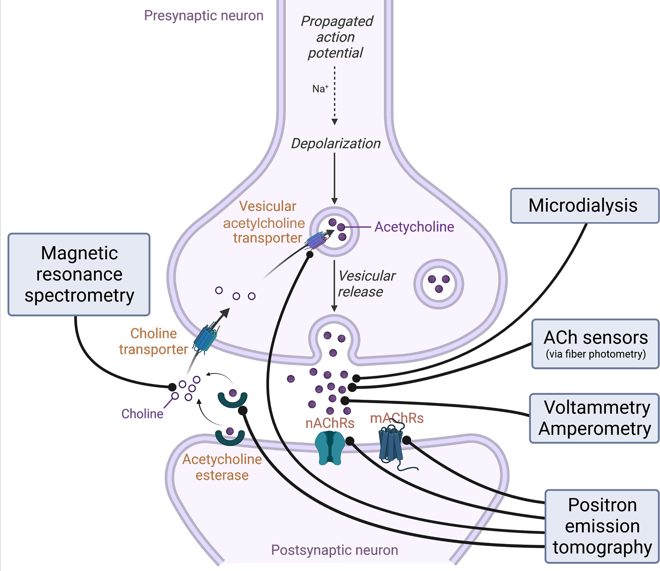
Figure 1: Schematic representation of an ACh release site and the targets for in vivo recording of ACh levels and cholinergic activity.

An example presynaptic cholinergic terminal is diagrammed, along with a generic postsynaptic cholinoceptive cell, with labeling of targets for several available methods of measuring ACh levels and activity. Positron emission tomography (PET) involves infusion of radioalabeled ligands that tag ACh binding sites followed by scanning, allowing estimation of various proxies for ACh levels and activity. Of note, some of these probes can compete for binding of ACh, and thus binding availability may represent either changes in levels of the binding site or changes in levels of endogenous ACh. Magnetic resonance imaging (MRS) can be used to detect the specific resonance spectrum of choline, a precursor of ACh whose levels are linearly related to ACh concentration and are altered in response to ACh release. Microdialysis samples fluid in the extracellular space and ACh levels are subsequently quantified using biochemical methods. Fiber photometry (FP) can be used to detect fluorescence changes due to calcium in the presynaptic terminal, as a proxy for activity of the cholinergic neurons, or as a result of ACh binding to fluorescence-based ACh sensors. Voltammetry and amperometry are used to detect specific electrochemical signatures from choline or ACh in the extracellular space.
