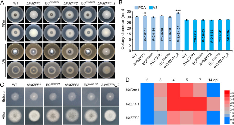
Fig. 2.
VdZFP1 and VdZFP2 play key roles in melanin production of Verticillium dahliae. A Colony morphology of WT, ΔVdZFP1, ECΔVdZFP1, ΔVdZFP2, ECΔVdZFP2, and ΔVdZFP1_2 inoculated on PDA and V8 media at 25 °C in the dark. The phenotypes were photographed 7 days after incubation. Each strain was inoculated at least three plates and three independent experiments were carried out. B Colony diameters of the different strains in panel A. Error bars are standard errors calculated from six replicates, and this experiment performed three repeats. *P < 0.05, **P < 0.01 (Student’s t test). C The penetrated ability of WT, ΔVdZFP1, ECΔVdZFP1, ΔVdZFP2, ECΔVdZFP1, and ΔVdZFP1_2 on cellophane membranes. The hyphal blocks were cut from PDA plates and placed on MM medium covered with cellophane membranes at 25 °C in the dark. The cellophane membranes were removed at 3 dpi, and the treated plates were incubated for an additional 5 days. This experiment performed three repeats. D Expression profile of VdZFP1, VdZFP2, and VdCmr1 during microsclerotia development of V. dahliae. The conidia of WT strain were grown on BMM medium covered with cellophane membranes at 25 °C in the dark and samples were collected at 2, 3, 4, 5, 7, and 14 dpi. The relative expression of VdZFP1, VdZFP2 and VdCmr1 were calculated from the RT-qPCR results using the 2.−ΔΔCT method with 2 dpi as control. This experiment was independently repeated 3 times to determine the trend. The results are presented in a heatmap (the expression levels were normalized to Log2 (fold-change))

