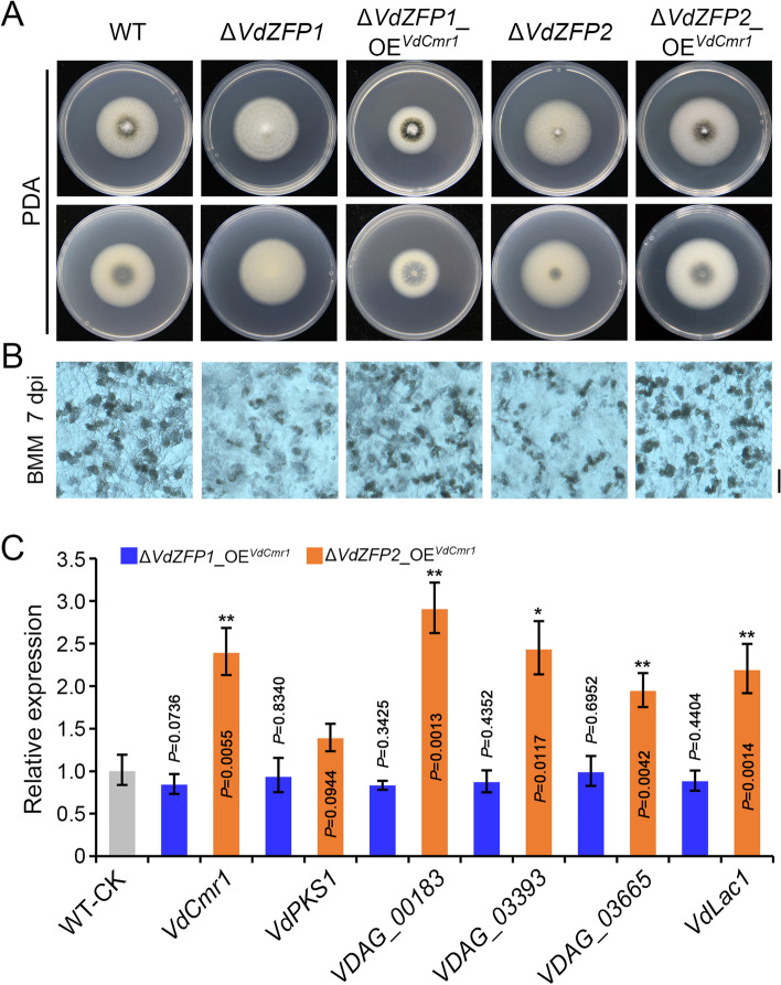
Fig. 6.
VdZFP1 and VdZFP2 mediate melanin deposition of microsclerotia by positively controlling VdCmr1 in Verticillium dahliae. A Colony morphology of the WT strain, mutants (ΔVdZFP1 and ΔVdZFP2), and VdCmr1-overexpressed strain (ΔVdZFP1_OEVdCmr1 and ΔVdZFP2_OEVdCmr1). The indicated strains were cultured on PDA medium at 25 °C in the dark and were photographed 7 days after incubation. Each strain was inoculated at least three plates. B Microsclerotia morphology of indicated strains. The microsclerotia development of each strain was observed at 7 dpi after incubating on the BMM plates covered with cellophane membranes at 25 °C in the dark and photographed with a stereoscope. Each strain was repeated three times independently, and at least three plates were observed each time. Scale bar = 100 μm. C Relative expression analyses of melanin-related genes in indicated strains. The strains were grown on BMM medium at 25 °C in the dark and were collected at 7 dpi for RT-qPCR. The results of 3 repetitions analyzed by the 2−ΔΔCT method with WT strain as control. This experiment was independently repeated 3 times to determine the trend. Error bars represent standard errors of the mean, and **P < 0.01, and.***P < 0.001 (one-way ANOVA)

