Fig. 7.
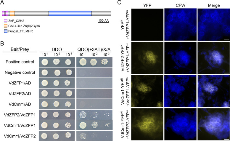
Verticillium dahliae VdZFP1, VdZFP2, and VdCmr1 interact directly with each other on the cell wall of microsclerotia precursors. A The conserved domains of VdCmr1 in V. dahliae. The protein sequence of VdCmr1 was predicted by SMART, InterPro, and Pfam and each domain was labeled in different colors. Scale bar = 100 amino acids. B Interaction analyses among VdZFP1, VdZFP2, and VdCmr1 in a yeast two-hybrid system. The CDS regions of VdZFP1, VdZFP2, and VdCmr1 were linked into pGADT7 and pGBKT7 vectors to obtain the prey and bait constructs. Each bait construct was co-transformed with pGADT7 vector into yeast cells to detect self-activation, while the yeast cells containing the bait and prey constructs were used to detect interaction. Yeast cells with tenfold serial dilutions were cultured on DDO (SD lacking Leu and Trp) medium and QDO (SD lacking Leu, Trp, His, and Ade) medium supplemented with 3AT (2.5 mM for pGBKT7-VdZFP1 and 70 mM for pGBKT7-VdCmr1), 20 μg/mL X-a-Gal, and 0.1 μg/mL AbA. The yeast cells co-transformed with pGADT7-T and pGBKT7-53 or pGBKT7-Lam vectors were set as the positive or negative control. The Interaction phenotypes were photographed at 5 dpi. This experiment was repeated 3 times. C Bimolecular fluorescence complementation assays among VdZFP1, VdZFP2, and VdCmr1. The recombinant plasmids of VdZFP1, VdZFP2, and VdCmr1 fused with C- or N-terminal of YFP were paired and co-transformed into WT strain. The recombinant plasmids co-transformed with YFPC or YFPN were negative controls. The CFW dye was used as the cell wall marker. The CFW and YFP signals were observed by fluorescence microscopy. Scale bars = 20 μm

