Fig 1.
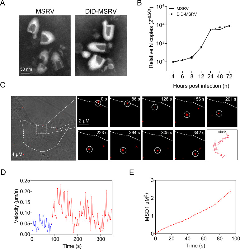
Characterization of labeled MSRV and real-time tracking of MSRV. (A) Transmission electron microscope images of unlabeled MSRV (left) and DiD (1,1′-dioctadecyl-3,3,3′,3′-tetramethylindodicarbocyanine,4-chlorobenzenesulfonate salt) labeled MSRV (right). Scale bars represent 50 nm. (B) Viral infection of DiD-MSRV in epithelioma papulosum cyprinid (EPC) cells. Cells were infected with MSRV at a multiplicity of infection (MOI) of 0.1. Viral RNA copy number was determined by RT-qPCR at 4, 6, 8, 12, 24, 48, and 72 hpi. (C) Confocal images of DiD-MSRV in MSRV infected EPC cells. White circles indicate the position of the virus. Dashed lines represent the plasma membrane. Red lines indicate the typical trajectories of virus. Scale bar = 2 µm. (D) Instantaneous velocity of the virus. Blue and red lines separately represent the slow and rapid movement of virus. (E) Mean-squared displacement (MSD)-time plots of viral movement.

