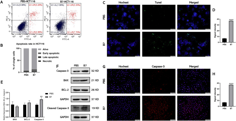
Figure 4.
B7 induces apoptosis of CRC cells by promoting the expression of Caspase-3. (A) Flow cytometry was used to analyze the effect of B7 on HCT116 cell apoptosis. (B) Statistical analysis of the percentage of HCT116 cells undergoing apoptosis. Statistical significance be-tween groups were evaluated using the Student’s t-test for independent groups. (C) TUNEL analysis of HCT116 cells treated with B7 (scale bar = 100 µm). And associated (D) statistical analysis of TUNEL fluorescence signal intensity. (E) Relative gene expression of apoptosis-related genes in HCT116 cells treated with PBS or B7. (F) Protein levels of BCL-2, BAX, Caspase-3, cleaved Caspase-3 and GAPDH in HCT116 cells treated with PBS or B7. (G) The fluorescent images of Caspase-3 in HCT116 cells treated with PBS or B7 (scale bar = 100µm). And associated (H) statistical analysis of the Caspase-3 intensity. The data are represented as the mean ± SD (n = 3). Statistical differences were considered significant at **p < 0.01 and ****p < 0.0001.

