
Fig 3.
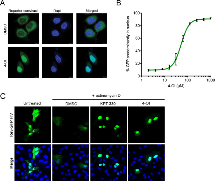
Impact of 4-OI on CRM1-dependent proteins. (A) The effect of treatment with 4-OI in a reporter HeLa cell line stably expressing a CRM1-dependent GFP reporter protein. Cells were treated with 100 µM 4-OI or DMSO for 24 hours, and the subcellular location was assessed by confocal microscopy. Confocal images are representative of three independent experiments. (B) Quantification of results from panel A. Cells were treated with indicated concentrations of 4-OI for 24 hours, and the percentage of cells with a predominantly nuclear localization of the GFP reporter protein was assessed. The experiment was conducted three times, and results are depicted as mean ± SD. (C) Confocal microscopy image of Vero-118 cells transfected with FIV Rev-GFP (green). Cells were left untreated or treated with actinomycin D simultaneously to DMSO, 100 µM 4-OI, or 100 nM KPT-330. Merge images include nuclear staining with Hoechst (blue). Confocal images are representative of three individual experiments.