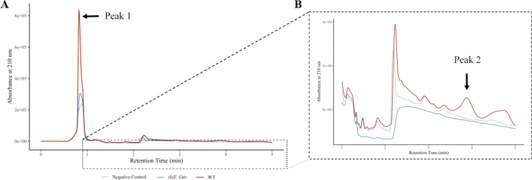
Fig 4.
HPLC analysis and UV detection. The pre-purified supernatant of the WT strain (red), rhiE::Gm mutant (blue) and non-inoculated medium (gray) were monitored at 210 nm after HPLC separation using a Milli-Q water/acetonitrile + 0.2% trifluoroacetic acid gradient for 25 min at 1 mL min−1. To determine the presence of siderophore activity, fractions were collected during the HPLC run and tested using the liquid CAS assay. Positive fractions for siderophore presence are highlighted with black arrows. (A) A major peak (peak 1) at retention time of 0.84 min was identified. (B) A second peak (peak 2) of lower intensity, with a retention time of 3.88 min, was identified after CAS test.

