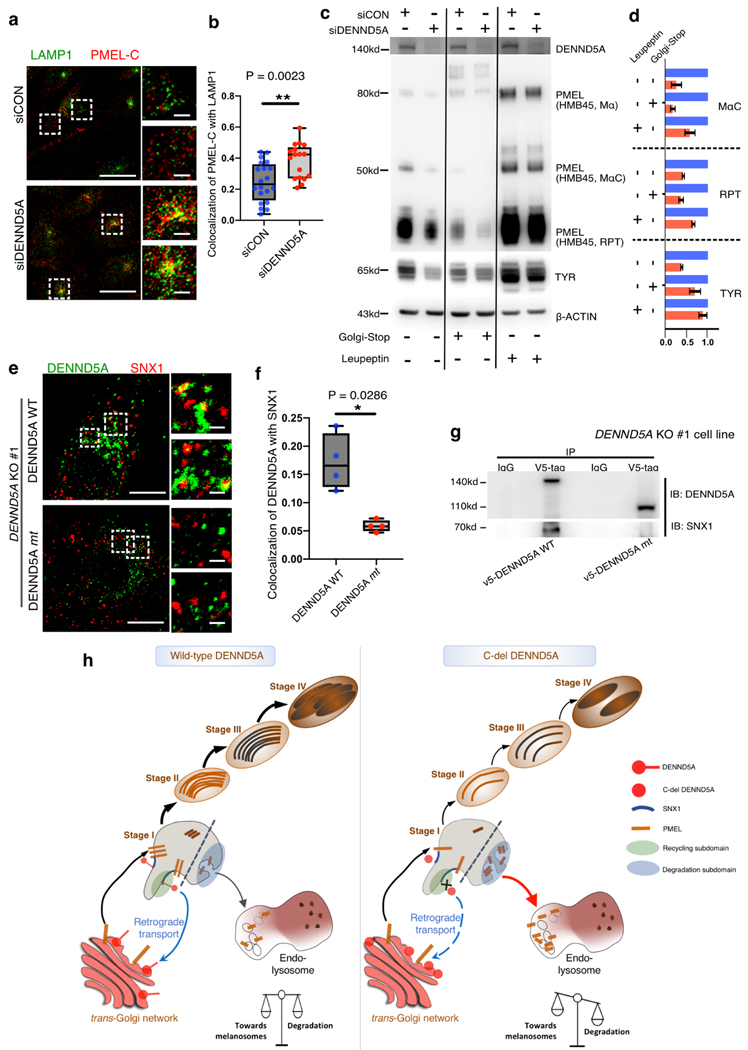
Fig. 4. Defective DENND5A triggers lysosomal degradation of melanosomal cargoes and fails to associate with SNX1-decorated vesicles.
a. Representative confocal microscopy images of control MNT-1 (siCON, upper) and after silencing DENND5A (siDENND5A, lower). LAMP1 (green) and PMEL (PMEL-C antibody, red) labeled MNT-1 cells are labeled and shown in merged channels and with higher magnification on the right-hand side. Co-localization is measured using images from two independent experiments and shown in b. Western blotting (c) and quantification (d) of melanosome markers for control (siCON)-MNT1 and siDENND5A-MNT1 after treatment of Golgi-Stop or lysosome protease inhibitor Leupeptin, respectively. e. DENND5A KO #1-MNT-1 cells were transiently introduced with V5-DENND5AWT (upper) and V5-DENND5Amt (lower) respectively. Transfected cells are stained for DENND5A (green) and SNX1 (red). Co-localization co-efficiency is measured using images from two independent experiments and shown in (f). Scale bars: 10 μm (a, e), 1 μm for the magnified panels. Data quantification is presented as mean ± S.E.M. Two-tailed, Unpaired t test (d), Mann-Whitney test (b, f). g. Immunoprecipitation of V5-tag on DENND5A KO #1 cells over-expressing the wild-type (V5-DENND5AWT) and mutated (V5-DENND5Amt) constructs. h. Proposed working model showing (left) the homeostasis of melanosome maturation and lysosomal degradation coordinated by DENND5A-SNX1 interaction. (right) C-del DENND5A loses its interaction with SNX1 and triggers mis-sorting of PMEL from en route to melanosomes to degradation subdomain (stage I melanosome)26, resulting in lysosomal entry and degradation of PMEL.

