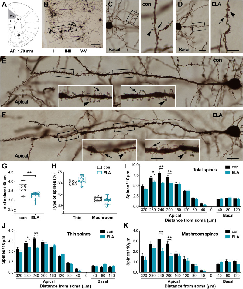
Fig. 1. ELA-provoked loss of spines on pyramidal cells in layers II-III of the prelimbic frontal cortex (PrL).
A, B Golgi-stained cells in the PrL. The representative image (B) was taken from a control mouse. Boxed areas in B were magnified in C and E to present the basal and apical dendrites of a pyramidal cell in layers II-III, respectively. IL: infralimbic; fmi: forceps minor of the corpus callosum; ac: anterior commissure. C, D Basal dendrites of pyramidal cells in layers II-III from a control and an ELA mouse. The boxed segments were magnified in the right panels to show thin (arrows) and mushroom-type (arrowheads) spines. E, F Apical dendrites of PrL pyramidal cells in a control and an ELA mouse. The boxed segments were magnified in insets to display numerous thin (arrows) and mushroom-type (arrowheads) spines in the control mouse and loss of spines in the ELA mouse. G–K Quantitative analysis. A decreased spine density was apparent in ELA mice (n = 9) versus controls (n = 8) (**P < 0.01). The percentage of thin and mushroom-type spines was not affected (H). ELA-provoked loss of spines (I) including both thin (J) and mushroom-type (K) spines was primarily observed on apical dendrites that were 200–280 µm from the soma of pyramidal cells (*P < 0.05, **P < 0.01). No difference was observed on basal dendrites. Scale bars = 60 µm (B), 15 µm (left panels in C, D and E, F), and 6 µm (right panels in C, D and insets in E, F).

