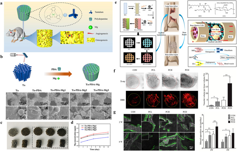
Fig. 3.
Metal-based and drug modification approaches for vascularized 3D printed networks. a Schematic illustration of metal ions modification onto the 3D printed scaffolds. b Schematic presentation of the fabrication process and SEM of the samples. c, d Optical images and cumulative release profile of metal ions. a–d are reproduced with permission [96].
Copyright 2020, Elsevier. e Schematic diagram of DFO on the surface of 3D printed PCL scaffold and its biological function for bone regeneration in bone defect model. f Typical X-ray photography and digitally reconstructed radiograph (DRR) after micro-CT scanning of the new vascular formed inside the bone defect site. g Dynamic histology of the newly formed bone using double calcein labeling after scaffolds insertion and the summarized data showing the difference of the mineral appositional rate among different groups. e–g are reproduced with permission [103]. Copyright 2018, Elsevier

