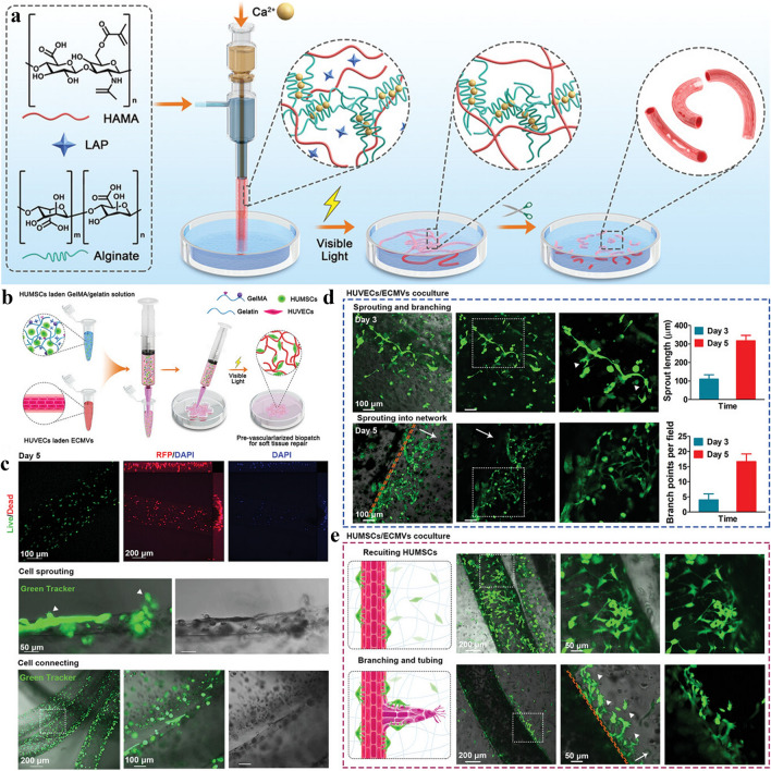
Fig. 6.
The cell-based and gene-based bioactive 3D printing vascular networks. a Schematic view of the printing process with cell-based bioink and microstructure images of the printed scaffolds. b Schematic diagram of the preparation of a repair hydrogel vascular scaffold containing human umbilical cord mesenchymal stem cells (HUMSC). c Live-dead staining to test the effectiveness of hydrogel vascular scaffolds for sprouting. d, e Confocal microscopy images tracing hydrogel vascular scaffold germination length and branching points over time. White triangular arrows indicate tip cells. While the long arrows indicate the invasion the direction of the newly formed network. a–e are reproduced with permission [190].
Copyright 2022, Wiley

