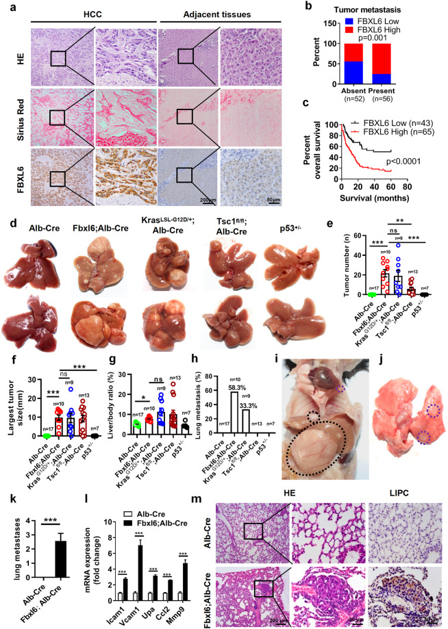
Fig. 1. FBXL6 is an independent risk factor for aggressive HCC and drives HCC lung metastasis in vivo significantly more strongly than Kras mutation, p53 loss or Tsc1 loss.
a Histologic analysis hematoxylin and eosin (H&E) and Sirius red staining and evaluation of FBXL6 expression by IHC staining in liver cancer tissues and adjacent normal tissues from 108 patients. Scale bars, 200 μm or 50 μm. b Association between FBXL6 protein expression and metastasis. c Association between FBXL6 protein expression and overall survival in HCC patients. The data are presented as the means ± SEMs. The log-rank (Mantel‒Cox) test was used, p < 0.0001. d–h Alb-Cre (n = 17), Fbxl6;Alb-Cre (n = 10), KrasLSL-G12D/+;Alb-Cre (n = 9), Tsc1fl/fl;Alb-Cre (n = 13), and p53+/– (n = 7) female mice were monitored for 310 days and were then euthanized. The livers were imaged (d). The liver tumor number (e), largest tumor size (f), liver/body weight ratio (g), and rate of lung metastasis (h) were analyzed. i, j Representative images showing lung metastasis in 310-day-old Fbxl6;Alb-Cre mice. k The number of observed metastases in mice of each genotype was quantified. l qPCR results showing the elevated expression of the metastasis markers Icam1, Vcam1, Upa, Ccl2, and Mmp9 in liver tumors of Fbxl6;Alb-Cre mice. n = 6. m Representative H&E and IHC staining images showing distant lung metastatic foci expressing the hepatocytic marker LIPC. Scale bars, 200 μm or 50 µm. The data are presented as the means ± SEMs. One-way ANOVA with the Bonferroni correction for multiple comparisons was used in (e–g); two-way ANOVA with the Bonferroni correction for multiple comparisons was used in (l); two-tailed unpaired t test was used in (k). ns nonsignificant. *p ≤ 0.05, **p ≤ 0.01, ***p ≤ 0.001.

