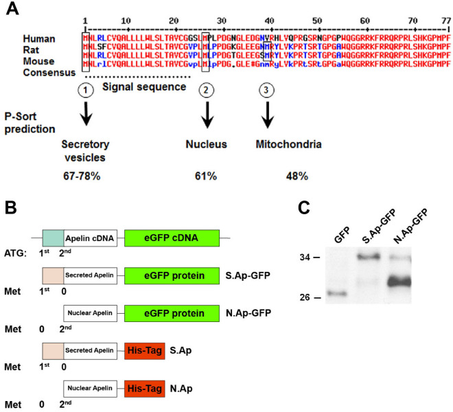
Figure 5.
Apelin translation at first or second initiation site. (A) Alignment of human, rat, and mouse preproApelin amino acid sequences and consensus sequence showing initiation sites at position 1 (①), 26 (②) and 39 (③). P-sort prediction for translation initiation starting at the first, second and third initiation site leads to signal sequences targeting proapelin to secretory vesicles, and shorter proapelin fragments to the nucleus and mitochondria, respectively. (B) Scheme showing point mutations generated at the first or second ATG site to abolish translation initiation with eGFP or His-Tagged apelin. (C) Western blot analysis of protein extracts 24 h after transfection of empty pEGFP-N1 (GFP), both mutated preproapelin encoded pEGFP-N1 (S. Ap-GFP and N. Ap-GFP) plasmids that were detected with GFP. GFP and fusion proteins are detected at the expected molecular weight. The figure represents a cropped image retaining important bands, original blots are presented in page 13 of the supplementary file.

