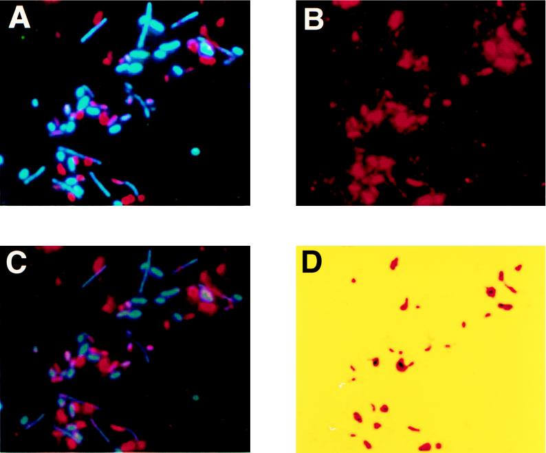
FIG. 2.
Detection and image analysis of E. coli in the model community. The ECOL primer was used in direct in situ PCR. (A) UV excitation image; (B) B excitation image; (C) composite image of UV and B images; (D) result of image analysis. Only target cells were identified. For an explanation, see the text. The final image analysis (D) was colored with pseudocolor.

