. 2023 Aug 8;13(11):2412–2431. doi: 10.1158/2159-8290.CD-23-0669

Figure 1.

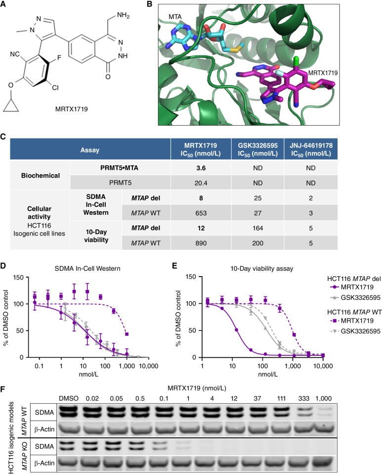
Figure 1. MRTX1719 binds PRMT5/MTA and selectively inhibits MTAP del cancer cells with reduced activity in MTAP WT cells. A, Chemical structure of MRTX1719. B, X-ray crystal structure of MRTX1719 cocomplexed with PRMT5/MEP50 and MTA (right; Protein Data Bank code 7S1S; ref. 14). C, MRTX1719 was run in a PRMT5/MEP50 biochemical assay that measures the activity of the complex in the absence and presence of MTA at an approximate IC50 concentration of MTA in the assay (2 μmol/L). MRTX1719, GSK3326595, and JNJ-64619178 were run in SDMA In-Cell Western (SYM11 antibody; C and D) and 10-day viability assays (C and E) in MTAP del and WT HCT116 cell lines. ND, not determined. F, Cropped SDMA Western blot showing protein bands that correlate with the molecular weight of SmD3 in MTAP del and WT HCT116 cell lines following 4 days of treatment with a range of concentrations of MRTX1719, with β-actin run as a loading control. KO, knockout.

MRTX1719 binds PRMT5/MTA and selectively inhibits MTAP del cancer cells with reduced activity in MTAP WT cells. A, Chemical structure of MRTX1719. B, X-ray crystal structure of MRTX1719 cocomplexed with PRMT5/MEP50 and MTA (right; Protein Data Bank code 7S1S; ref. 14). C, MRTX1719 was run in a PRMT5/MEP50 biochemical assay that measures the activity of the complex in the absence and presence of MTA at an approximate IC50 concentration of MTA in the assay (2 μmol/L). MRTX1719, GSK3326595, and JNJ-64619178 were run in SDMA In-Cell Western (SYM11 antibody; C and D) and 10-day viability assays (C and E) in MTAP del and WT HCT116 cell lines. ND, not determined. F, Cropped SDMA Western blot showing protein bands that correlate with the molecular weight of SmD3 in MTAP del and WT HCT116 cell lines following 4 days of treatment with a range of concentrations of MRTX1719, with β-actin run as a loading control. KO, knockout.